

Published : 28 Sep 2026, 06:55 PM
Updated : 28 Sep 2026, 06:55 PM


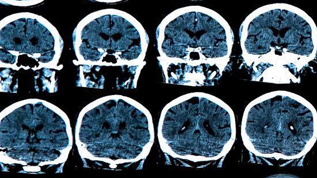
MRI scans could help researchers understand how Alzheimer’s disease develops years before its better-known markers appear, according to Science News.
Researchers found that people who later developed high levels of amyloid-beta had slightly thicker cortices than those who did not. The differences, measured in hundredths of a millimetre, were visible up to seven years before high amyloid-beta levels were detected through PET scans.
The cortex, the brain’s outermost layer, normally becomes thinner with age. However, a study published in Nature Neuroscience found that it thinned more slowly in people who later developed high amyloid-beta levels.
The researchers did not find a link between the early cortical changes and memory problems.
According to Science News, findings suggest that changes in the brain during the early stages of Alzheimer’s may be more complex than previously thought. Victor Montal, a degenerative diseases researcher at the Barcelona Supercomputing Center, said the “trajectory of cortical changes is not as simplistic as we used to think”.
The researchers said inflammation linked to the early accumulation of amyloid-beta could explain the temporary thickening, although MRI scans cannot establish the cause directly.
The team analysed MRI scans from more than 1,000 participants, with the cortex averaging about 2.5 millimetres thick.
Science News said the changes are currently too subtle to serve as an individual diagnostic tool. Frederik Barkhof, a neuroradiologist at University College London, said, “I don’t think that this cortical thickness is something you’re going to use clinically.”
However, the findings could help scientists better understand Alzheimer’s mechanisms and assess how treatments affect changes in the brain. Some existing drugs remove amyloid-beta but do not prevent brain volume loss, and the new findings could offer clues about the role of inflammation in that process.